
Acute respiratory distress syndrome ("ARDS"), previously known as respiratory distress syndrome ("RDS"), adult respiratory distress syndrome, or shock lung, is a severe, life-threatening medical condition characterized by widespread inflammation in the lungs. Although it can be triggered by a respiratory infection, such as pneumonia, it is more often a result of sepsis or significant trauma.
ARDS is a disease of the lung parenchyma that leads to impaired gas exchange. It is associated with pulmonary cytokine release, impaired endothelial barriers, loss of surfactant, fluid accumulation in the distal airspaces, and fibrotic changes. The mortality rate varies widely based on disease severity and patient age, with an average mortality somewhere between 20% and 50%.
The acronym ARDS formerly signified "adult respiratory distress syndrome" to differentiate it from "infant respiratory distress syndrome", which occurs in premature infants. However, as this type of pulmonary edema also occurs in children, ARDS has gradually shifted to mean "acute" rather than "adult". The differences from the typical infant syndrome remain.
Signs and symptoms
ARDS is an acute injury to the lungs that results in alveolar flooding, atelectasis and a severe oxygenation defect, but is not due to heart failure. In its early stages, it can cause shortness of breath (dyspnea), increased rate of breathing (tachypnea), and low oxygen saturation (hypoxemia).
ARDS is characterized by the following criteria:
- lung injury of acute onset, within 1 week of an apparent clinical insult and with progression of respiratory symptoms
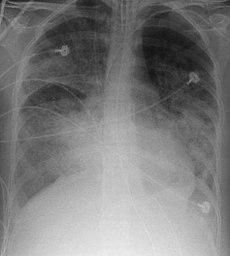
- bilateral opacities on chest imaging not explained by other pulmonary pathology (e.g. pleural effusion, pneumothorax, or nodules)
- respiratory failure not explained by heart failure or volume overload
- decreased arterial PaO
2/FiO
2 ratio:- mild ARDS: ratio is 201 - 300 mmHg (≤ 39.9 kPa)
- moderate ARDS: 101 - 200 mmHg (≤ 26.6 kPa)
- severe ARDS: ≤ 100 mmHg (≤ 13.3 kPa)
(a minimum PEEP of 5Â cmH
2O is required; it may be delivered noninvasively with CPAP to diagnose mild ARDS). A decreased PaO
2/FiO
2 ratio indicates reduced arterial oxygen content relative to that of the inhaled gas, indicating a failure of the lung to transport oxygen into the blood.
The above characteristics are the "Berlin criteria" of 2012 by the European Society of Intensive Care Medicine, endorsed by the American Thoracic Society and the Society of Critical Care Medicine. They are a modification of the previously used criteria:
- Acute onset
- Bilateral infiltrates on chest radiograph sparing costophrenic angles
- Pulmonary artery wedge pressure < 18 mmHg (obtained by pulmonary artery catheterization), if this information is available; if unavailable, then lack of clinical evidence of left atrial hypertension
- if PaO
2:FiO
2 < 300 mmHg (40 kPa) acute lung injury (ALI) is considered to be present - if PaO
2:FiO
2 < 200 mmHg (26.7 kPa) acute respiratory distress syndrome (ARDS) is considered to be present
Cause
The predisposing factors of ARDS are numerous and varied. Sepsis, multiple blood transfusions, pulmonary contusion, aspiration of gastric contents and drug abuse or overdose are common. Also, burns, pancreatitis, smoke inhalation, pneumonia and near drowning can cause this condition. The inhalation of irritants, chemical warfare agents such as phosgene, chlorine gas and such can also cause ARDS.
Some cases of ARDS are linked to large volumes of fluid used during post-trauma resuscitation. Other causes include shock, near-drowning and inhalation of irritants or toxic fumes that damage the alveolar epithelium.
The list of predisposing factors is extensive and some do not necessarily seem to have anything to do with injuring the lungs. Therefore, this syndrome is best diagnosed and managed on its criteria first, with retrospective management on whatever conditions may have precipitated it.
Diagnosis

An arterial blood gas analysis and chest X-ray allow formal diagnosis. Although severe hypoxemia is generally included, the appropriate threshold defining abnormal PaO
2 has never been systematically studied. A severe oxygenation defect is not synonymous with ventilatory support. Any PaO
2 below 100 (generally saturation less than 100%) on a supplemental oxygen fraction of 50% meets criteria for ARDS. This can easily be achieved by high flow oxygen supplementation without ventilatory support. While CT scanning leads to more accurate images of the pulmonary parenchyma in ARDS, it has little use in the clinical management of patients with ARDS and remains largely a research tool.
Pathophysiology
ARDS is a clinical syndrome associated with pathological findings including pneumonia, eosinophilic pneumonia, cryptogenic organizing pneumonia, acute fibrinous organizing pneumonia, and diffuse alveolar damage (DAD). Of these, the pathology most commonly associated with ARDS is DAD, which is characterized by a diffuse inflammation of lung parenchyma. The triggering insult to the parenchyma usually results in an initial release of cytokines and other inflammatory mediators secreted by local epithelial and endothelial cells.
Neutrophils and some T-lymphocytes quickly migrate into the inflamed lung parenchyma and contribute in the amplification of the phenomenon. Typical histological presentation involves diffuse alveolar damage and hyaline membrane formation in alveolar walls. Although the triggering mechanisms are not completely understood, recent research has examined the role of inflammation and mechanical stress.
Inflammation
Inflammation, such as that caused by sepsis, causes endothelial dysfunction, fluid extravasation from the capillaries and impaired drainage of fluid from the lungs. Dysfunction of type II pulmonary epithelial cells may also be present, with a concomitant reduction in surfactant production. Elevated inspired oxygen concentration often becomes necessary at this stage, and may facilitate a 'respiratory burst' in immune cells. In a secondary phase, endothelial dysfunction causes cells and inflammatory exudate to enter the alveoli. This pulmonary edema increases the thickness of the alveolo-capillary space, increasing the distance the oxygen must diffuse to reach blood, which impairs gas exchange leading to hypoxia, increases the work of breathing and eventually induces fibrosis of the airspace.
Edema and decreased surfactant production by type II pneumocytes may cause whole alveoli to collapse or to completely flood. This loss of aeration contributes further to the right-to-left shunt in ARDS. As the alveoli contain progressively less gas, the blood flowing through the alveolar capillaries is progressively less oxygenated, resulting in massive intrapulmonary shunting. Collapsed alveoli and small bronchi do not allow gas exchange. It is common to see patients with a PaO
2 of 60 mmHg (8.0 kPa) despite mechanical ventilation with 100% inspired oxygen.
The loss of aeration may follow different patterns depending upon the nature of the underlying disease and other factors. In pneumonia-induced ARDS for example, large, more commonly causes relatively compact areas of alveolar infiltrates. These are usually distributed to the lower lobes, in their posterior segments, and they roughly correspond to the initial infected area. In sepsis or trauma-induced ARDS, infiltrates are usually more patchy and diffuse. The posterior and basal segments are always more affected, but the distribution is even less homogeneous. Loss of aeration also causes important changes in lung mechanical properties that are fundamental in the process of inflammation amplification and progression to ARDS in mechanically ventilated patients.
Mechanical stress
Mechanical ventilation is an essential part of the treatment of ARDS. As loss of aeration and the underlying disease progress, the end tidal volume grows to a level incompatible with life. Thus, mechanical ventilation is initiated to relieve respiratory muscles of their work and to protect the usually obtunded patient's airways. However, mechanical ventilation may constitute a risk factor for the developmentâ€"or the worseningâ€"of ARDS. Aside from the infectious complications arising from invasive ventilation with tracheal intubation, positive-pressure ventilation directly alters lung mechanics during ARDS. When these techniques are used the result is higher mortality through barotrauma.
In 1998, Amato et al. published a paper showing substantial improvement in the outcome of patients ventilated with lower tidal volumes (Vt) (6 mL·kgâˆ'1). This result was confirmed in a 2000 study sponsored by the NIH. Both studies were widely criticized for several reasons, and the authors were not the first to experiment with lower-volume ventilation, but they increased the understanding of the relationship between mechanical ventilation and ARDS.
One opinion is that the forces applied to the lung by the ventilator may work as a lever to induce further damage to lung parenchyma. It appears that shear stress at the interface between collapsed and aerated units may result in the breakdown of aerated units, which inflate asymmetrically due to the 'stickiness' of surrounding flooded alveoli. The fewer such interfaces around an alveolus, the lesser the stress. Even relatively low stress forces may induce signal transduction systems at the cellular level, thus inducing the release of inflammatory mediators.
This form of stress is thought to be applied by the transpulmonary pressure (gradient) (Pl) generated by the ventilator or, better, its cyclical variations. The better outcome obtained in patients ventilated with lower Vt may be interpreted as a beneficial effect of the lower Pl. Transpulmonary pressure is an indirect function of the Vt setting on the ventilator, and only trial patients with plateau pressures (a surrogate for the actual Pl) were less than 32 cmH
2O (3.1 kPa) had improved survival.
The way Pl is applied on alveolar surface determines the shear stress to which lung units are exposed. ARDS is characterized by a usually inhomogeneous reduction of the airspace, and thus by a tendency towards higher Pl at the same Vt, and towards higher stress on less diseased units. The inhomogeneity of alveoli at different stages of disease is further increased by the gravitational gradient to which they are exposed and the different perfusion pressures at which blood flows through them. Finally, abdominal pressure exerts an additional pressure on inferoposterior lung segments, favoring compression and collapse of those units.
The different mechanical properties of alveoli in ARDS may be interpreted as having varying time constantsâ€"the product of alveolar compliance × resistance. A long time constant indicates an alveolus which opens slowly during tidal inflation, as a consequence of contrasting pressure around it, or altered water-air interface inside itâ€"loss of surfactant, flooding.
Slow alveoli are said to be "kept open" using positive end-expiratory pressure, a feature of modern ventilators which maintains a positive airway pressure throughout the whole respiratory cycle. A higher mean pressure cycle-wide slows the collapse of diseased units, but it has to be weighed against the corresponding elevation in Pl/plateau pressure. Newer ventilatory approaches attempt to maximize mean airway pressure for its ability to "recruit" collapsed lung units while minimizing the shear stress caused by frequent openings and closings of aerated units. The prone position also reduces the inhomogeneity in alveolar time constants induced by gravity and edema. If clinically appropriate, mobilization of the ventilated patient can assist in achieving the same goal.
Stress Index
Mechanical ventilation can exacerbate the inflammatory response in patients with ARDS by including cyclic tidal alveolar hyperinflation and/or recruiting/derecruiting. Stress index is measured during constant-flow assist-control mechanical ventilation without changing the baseline ventilatory pattern. Identifying the steadiest portion of the inspiratory flow (F) waveform fit the corresponding portion of the airway pressure (Paw) waveform in the following power equation:
Paw = a × tb + c where the coefficient bâ€"the Stress Indexâ€"describes the shape of the curve. The Stress Index depict a constant compliance if the value is around 1, an increasing compliance during the inspiration if the value is below 1, and a decreasing compliance if the value is above 1. Ranieri, Grasso, et al. set a strategy guided by the stress index with the following rules:
- Stress Index below 0.9, PEEP was increased
- Stress Index between 0.9 and 1.1, no change was made
- Stress Index above 1.1 PEEP was decreased.
- Adjustment of PEEP was suspended if any one of the following conditions ensued: plateau pressure > 30Â cmH
2O, SaO
2 < 88%, or hemodynamic instability.
Alveolar hyperinflation in patients with focal ARDS ventilated with the ARDSnet protocol is attenuated by a physiologic approach to PEEP setting based on the stress index measurement.
Progression
If the underlying disease or injurious factor is not removed, the quantity of inflammatory mediators released by the lungs in ARDS may result in a systemic inflammatory response syndrome or sepsis if there is lung infection. The evolution towards shock or multiple organ failure follows paths analogous to the pathophysiology of sepsis. This leads to the impaired oxygenation which is the central problem of ARDS, as well as to respiratory acidosisâ€"which is often caused by ventilation techniques such as permissive hypercapnia, which attempt to limit ventilator-induced lung injury in ARDS. The result is a critical illness in which the 'endothelial disease' of severe sepsis or SIRS is worsened by the pulmonary dysfunction, which further impairs oxygen delivery.
Treatment
Acute respiratory distress syndrome is usually treated with mechanical ventilation in the Intensive Care Unit. Ventilation is usually delivered through pro-tracheal intubation, or by tracheotomy whenever prolonged ventilation (≥2 weeks) is deemed inevitable. The possibilities of non-invasive ventilation are limited to the very early period of the disease or to prevention in individuals with atypical pneumonias, pulmonary contusion, or major surgery patients, who are at risk of developing ARDS. Treatment of the underlying cause is imperative. Appropriate antibiotic therapy must be administered as soon as microbiological culture results are available. Empirical therapy may be appropriate if local microbiological surveillance is efficient. More than 60% ARDS patients experience a (nonsocial) pulmonary infection either before or after the onset of lung injury.
The origin of infection, when surgically treatable, must be operated on. When sepsis is diagnosed, appropriate local protocols should be enacted. Commonly used supportive therapy includes particular techniques of mechanical ventilation and pharmacological agents whose effectiveness with respect to the outcome has not yet been proven. It is now debated whether mechanical ventilation is to be considered mere supportive therapy or actual treatment, since it may substantially affect survival. Survivors of ARDS have an increased risk of lower quality of life, persistent cognitive impairment, depression and post-traumatic stress disorder.
Mechanical ventilation
The overall goal is to maintain acceptable gas exchange and to minimize adverse effects in its application. The parameters PEEP (positive end-expiratory pressure, to maintain maximal recruitment of alveolar units), mean airway pressure (to promote recruitment and predictor of hemodynamic effects) and plateau pressure (best predictor of alveolar overdistention) are used.
Conventional therapy aimed at tidal volumes (Vt) of 12â€"15 ml/kg (where the weight is ideal body weight rather than actual weight). Recent studies have shown that high tidal volumes can overstretch alveoli resulting in volumetric (secondary lung injury). The ARDS Clinical Network, or ARDSNet, completed a trial that showed improved mortality when ventilated with a tidal volume of 6 ml/kg compared to the traditional 12 ml/kg. Low tidal volumes (Vt) may cause hypercapnia and atelectasis because of their inherent tendency to increase physiologic shunt. Physiologic dead space cannot change as it is ventilation without perfusion. A shunt is perfusion without ventilation.
Low tidal volume ventilation was the primary independent variable associated with reduced mortality in the NIH-sponsored ARDSnet trial of tidal volume in ARDS. Plateau pressure less than 30Â cm H
2O was a secondary goal, and subsequent analyses of the data from the ARDSnet trial and other experimental data demonstrate that there appears to be no safe upper limit to plateau pressure; regardless of plateau pressure, patients fare better with low tidal volumes.
Airway pressure release ventilation
It is often said that no particular ventilator mode is known to improve mortality in airway pressure release ventilation (APRV).
Some practitioners favor airway pressure release ventilation when treating ARDS. Well documented advantages to APRV ventilation include: decreased airway pressures, decreased minute ventilation, decreased dead-space ventilation, promotion of spontaneous breathing, almost 24 hour a day alveolar recruitment, decreased use of sedation, near elimination of neuromuscular blockade, optimized arterial blood gas results, mechanical restoration of FRC (functional residual capacity), a positive effect on cardiac output (due to the negative inflection from the elevated baseline with each spontaneous breath), increased organ and tissue perfusion and potential for increased urine output secondary to increased renal perfusion.
A patient with ARDS, on average, spends between 8 and 11 days on a mechanical ventilator; APRV may reduce this time significantly and conserve valuable resources.
Positive end-expiratory pressure
Positive end-expiratory pressure (PEEP) is used in mechanically ventilated patients with ARDS to improve oxygenation. In ARDS, three populations of alveoli can be distinguished. There are normal alveoli which are always inflated and engaging in gas exchange, flooded alveoli which can never, under any ventilatory regime, be used for gas exchange, and atelectatic or partially flooded alveoli that can be "recruited" to participate in gas exchange under certain ventilatory regimens. The recruitable aveoli represent a continuous population, some of which can be recruited with minimal PEEP, and others which can only be recruited with high levels of PEEP. An additional complication is that some alveoli can only be opened with higher airway pressures than are needed to keep them open, hence the justification for maneuvers where PEEP is increased to very high levels for seconds to minutes before dropping the PEEP to a lower level. PEEP can be harmful; high PEEP necessarily increases mean airway pressure and alveolar pressure, which can damage normal alveoli by overdistension resulting in DAD. A compromise between the beneficial and adverse effects of PEEP is inevitable.
The 'best PEEP' used to be defined as 'some' cmH
2O above the lower inflection point (LIP) in the sigmoidal pressure-volume relationship curve of the lung. Recent research has shown that the LIP-point pressure is no better than any pressure above it, as recruitment of collapsed alveoliâ€"and more importantly the overdistension of aerated unitsâ€"occur throughout the whole inflation. Despite the awkwardness of most procedures used to trace the pressure-volume curve, it is still used by some to define the minimum PEEP to be applied to their patients. Some new ventilators can automatically plot a pressure-volume curve.
PEEP may also be set empirically. Some authors suggest performing a 'recruiting maneuver'â€"a short time at a very high continuous positive airway pressure, such as 50 cmH
2O (4.9 kPa)â€"to recruit or open collapsed units with a high distending pressure before restoring previous ventilation. The final PEEP level should be the one just before the drop in PaO
2 or peripheral blood oxygen saturation during a step-down trial.
Intrinsic PEEP (iPEEP) or auto-PEEPâ€"first described by John Marini of St. Paul Regions Hospitalâ€"is a potentially unrecognized contributor to PEEP in patients. When ventilating at high frequencies, its contribution can be substantial, particularly in patients with obstructive lung disease. iPEEP has been measured in very few formal studies on ventilation in ARDS patients, and its contribution is largely unknown. Its measurement is recommended in the treatment of ARDS patients, especially when using high-frequency (oscillatory/jet) ventilation.
Prone position
Distribution of lung infiltrates in acute respiratory distress syndrome is non-uniform. Repositioning into the prone position (face down) might improve oxygenation by relieving atelectasis and improving perfusion. If this is done early in the treatment of severe ARDS, it confers a mortality benefit of 26% compared to supine ventilation.
Fluid management
Several studies have shown that pulmonary function and outcome are better in patients that lost weight or pulmonary wedge pressure was lowered by diuresis or fluid restriction.
Corticosteroids
A study found improvement in ARDS using modest doses of corticosteroids. The initial regimen consists of methylprednisolone. After 3â€"5 days a response must be apparent. In 1â€"2 weeks the dose can be tapered. But high dose steroid therapy has no effect on ARDS when given within 24 hours of the onset of illness. This study involved a small number of patients in one center. A recent NIH-sponsored multicenter ARDSnet LAZARUS study of corticosteroids for ARDS demonstrated that they are not efficacious in ARDS. The benefit of steroids in late ARDS may be explained by the ability of steroids to promote breakdown and inhibit fibrosis as well as decreases edema.
Nitric oxide
Inhaled nitric oxide (NO) potentially acts as selective pulmonary vasodilator. Rapid binding to hemoglobin prevents systemic effects. It should increase perfusion of better ventilated areas. There are no large studies demonstrating positive results; its use must be considered individually. Almitrine bismesylate stimulates chemoreceptors in carotic and aortic bodies. It has been used to potentiate the effect of NO, presumably by potentiating hypoxia-induced pulmonary vasoconstriction. In case of ARDS it is not known whether this combination is useful.
Surfactant therapy
To date no prospective controlled clinical trial has shown a significant mortality benefit of exogenous surfactant in adult ARDS.
Complications

Since ARDS is an extremely serious condition which requires invasive forms of therapy it is not without risk. Complications to be considered are:
- Pulmonary: barotrauma (volutrauma), pulmonary embolism (PE), pulmonary fibrosis, ventilator-associated pneumonia (VAP)
- Gastrointestinal: hemorrhage (ulcer), dysmotility, pneumoperitoneum, bacterial translocation
- Cardiac: arrhythmias, myocardial dysfunction
- Renal: acute renal failure (ARF), positive fluid balance
- Mechanical: vascular injury, pneumothorax (by placing pulmonary artery catheter), tracheal injury/stenosis (result of intubation and/or irritation by endotracheal tube
- Nutritional: malnutrition (catabolic state), electrolyte deficiency.
Epidemiology
The annual incidence of ARDS is 13-23 people per 100,000 in the general population. Its incidence in the mechanically ventilated population in intensive care units is much higher. According to Brun-Buisson et al (2004), there is a prevalence of acute lung injury (ALI) of 16.1% percent in ventilated patients admitted for more than 4 hours. More than half of these patients may develop ARDS.
ARDS may be triggered by mechanical ventilation, sepsis, pneumonia, Gilchrist's disease, circulatory shock, aspiration, traumaâ€"especially pulmonary contusionâ€"major surgery, massive blood transfusions, smoke inhalation, drug reaction or overdose, fat emboli and reperfusion pulmonary edema after lung transplantation or pulmonary embolectomy. Pneumonia and sepsis are the most common triggers, and pneumonia is present in up to 60% of patients and may be either causes or complications of ARDS. Alcohol excess appears to increase the risk of ARDS. Diabetes was originally thought to decrease the risk of ARDS, but this has shown to be due to an increase in the risk of pulmonary oedema.
Elevated abdominal pressure of any cause is also probably a risk factor for the development of ARDS, particularly during mechanical ventilation. The death rate varies from 25-40% in centers using up-to-date ventilatory strategies and up to 58% in all centers. Usually, randomized controlled trials in the literature show lower death rates than those shown by observational studies, both in control and treatment patients. This is thought to be due to stricter enrollment criteria.
History
Acute respiratory distress syndrome was first described in 1967 by Ashbaugh et al. Initially there was no definition, resulting in controversy over incidence and death. In 1988, an expanded definition was proposed which quantified physiologic respiratory impairment.
In 1994, a new definition was recommended by the American-European Consensus Conference Committee. It is simple to use and it recognizes that severity of pulmonary injury varies.
ARDS was defined as the ratio of arterial partial oxygen tension (PaO
2) as fraction of inspired oxygen (FiO
2) below 200 mmHg in the presence of bilateralinfiltrates on the chest x-ray. These infiltrates may appear similar to those of left ventricular failure, but the cardiac silhouette appears normal in ARDS. Also, the pulmonary capillary wedge pressure is normal (less than 18 mmHg) in ARDS, but raised in left ventricular failure. A PaO
2/FiO
2 ratio less than 300 mmHg with bilateral infiltrates indicates acute lung injury (ALI). Although formally considered different from ARDS, ALI is usually a precursor to ARDS.
In 2012 the Berlin Definition of ARDS was published, removing the ALI/ARDS differentiation. It opts instead to classify ARDS as mild, moderate or severe.


COPD is a disease that affects the lung. This disease cause breathing problem and poor air flow from the lung. There are various types of treatment available for this treatment. But stem cell therapy is the most effective treatment for this disease. Stem cells COPD treatment
ReplyDeleteThis blog is so nice to me. I will keep on coming here again and again. Visit my link as well..
ReplyDeletes4