
An intracranial aneurysm (also called cerebral or brain aneurysm) is a cerebrovascular disorder in which weakness in the wall of a cerebral artery or vein causes a localized dilation or ballooning of the blood vessel.
Aneuryms in the posterior circulation (basilar, vertebral and posterior communicating arteries) have a higher risk of rupture. Basilar artery aneurysms represent only 3%-5% of all intracranial aneurysms but are the most common aneurysms in the posterior circulation.
Classification
Cerebral aneurysms are classified both by size and shape. Small aneurysms have a diameter of less than 15mm. Larger aneurysms include those classified as large (15 to 25mm), giant (25 to 50mm), and super giant (over 50mm).
Saccular aneurysms
Saccular aneurysms, also known as berry aneurysms, appear as a round outpouching and are the most common form of cerebral aneurysm.
Fusiform aneurysms
Fusiform dolichoectatic aneurysms represent a widening of a segment of an artery around the entire blood vessel, rather than just arising from a side of an artery's wall. They can rupture but usually do not.
Microaneurysms
Microaneurysms, also known as Charcot-Bouchard aneurysms, typically occur in small blood vessels (less than 300 micrometre diameter), most often the lenticulostriate vessels of the basal ganglia, and are associated with chronic hypertension. Charcotâ€"Bouchard aneurysms are a common cause of intracranial hemorrhage.
Signs and symptoms
A small, unchanging aneurysm will produce few, if any, symptoms. Before a larger aneurysm ruptures, the individual may experience such symptoms as a sudden and unusually severe headache, nausea, vision impairment, vomiting, and loss of consciousness, or the individual may be asymptomatic (i.e., experiencing no symptoms at all).
Subarachnoid haemorrhage
If an aneurysm ruptures, blood leaks into the space around the brain. This is called a subarachnoid hemorrhage. Onset is usually sudden with no prodrome, classically presenting as a "thunderclap headache" worse than previous headaches. Symptoms of a subarachnoid hemorrhage differ depending on the site and size of the aneurysm. Symptoms of a ruptured aneurysm can include:
- a sudden severe headache that can last from several hours to days
- nausea and vomiting
- drowsiness, confusion and/or loss of consciousness
- Visual abnormalities
- Meningism
The risk of a subarachnoid hemorrhage is greater with a saccular aneurysm than a fusiform aneurysm.
Microaneurysms
A ruptured microaneurysm may cause an intracerebral hemorrhage, presenting as a focal neurological deficit.
Rebleeding, hydrocephalus (the excessive accumulation of cerebrospinal fluid), vasospasm (spasm, or narrowing, of the blood vessels), or multiple aneurysms may also occur. The risk of rupture from an unruptured cerebral aneurysm varies according to the size of an aneurysm, with the risk rising as the aneurysm size increases.
Risk factors
Intracranial aneurysms may result from diseases acquired during life, or from genetic conditions. Lifestyle diseases including hypertension, smoking, excess alcohol consumption, and obesity are associated with the development of aneurysms. Cocaine use has also been associated with the development of intracranial aneurysms.
Other acquired associations with intracranial aneurysms include head trauma and infections.
Genetic associations
Coarctation of aorta is also a known risk factor, as may arteriovenous malformation.
Genetic conditions associated with connective tissue disease may also be associated with the development of aneurysms. This includes:
- autosomal dominant polycystic kidney disease,
- neurofibromatosis type I,
- Marfan syndrome,
- multiple endocrine neoplasia type I,
- pseudoxanthoma elasticum,
- hereditary hemorrhagic telangiectasia and
- Ehlers-Danlos syndrome type II and IV.
Specific genes have also had reported association with the development of intracranial aneurysms, including perlecan, elastin, collagen type 1 A2, endothelial nitric oxide synthase, endothelin receptor A and cyclin dependent kinase inhibitor - have been reported. Mutations in interleukin 6 may be protective.. Recently, several genetic loci have been identified as relevant to the development of intracranial aneurysms. These include 1p34-36, 2p14-15, 7q11, 11q25, and 19q13.1-13.3.
Pathophysiology

Aneurysm means an outpouching of a blood vessel wall that is filled with blood. Aneurysms occur at a point of weakness in the vessel wall. This can be because of acquired disease or hereditary factors. The repeated trauma of blood flow against the vessel wall presses against the point of weakness and causes the aneurysm to enlarge. As described by the Law of Young-Laplace, the increasing area increases tension against the aneurysmal walls, leading to enlargement.
Saccular aneurysms
Saccular aneurysms are almost always the result of hereditary weakness in blood vessels and typically occur within the arteries of the Circle of Willis, in order of frequency affecting the following arteries:
- Anterior communicating artery
- Posterior communicating artery
- Middle cerebral artery
- Internal carotid artery
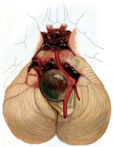
- Tip of basilar artery
Saccular aneurysms tend to have a lack of tunica media and elastic lamina around its dilated location (congenital), with wall of sac made up of thickened hyalinized intima and adventitia. In addition, some parts of the brain vasculature are inherently weakâ€"particularly areas along the Circle of Willis, where small communicating vessels link the main cerebral vessels. These areas are particularly susceptible to saccular aneurysms. Approximately 25% patients have multiple aneurysms, predominantly when there is familial pattern.
Complications

Rupture
Almost all aneurysms rupture at their apex. This leads to hemorrhage in the subarachnoid space and sometimes in brain parenchyma. Minor leakage from aneurysm may precede rupture, causing warning headaches. About 60% of patients die immediately after rupture. Larger aneurysms have greater tendency to rupture, though most of ruptured aneurysms are less than 10Â mm in diameter.
Vasospasm
Vasospasm, referring to blood vessel constriction, can occur secondary to subarachnoid hemorrhage following a ruptured aneurysm. This is most likely to occur within 21 days and is seen radiologically within 60% of such patients. The vasospasm is thought to be secondary to the apoptosis of inflammatory cells such as macrophages and neutrophils that become trapped in the subarachnoid space. These cells initially invade the subarachnoid space from the circulation in order to phagocytose the hemorrhaged red blood cells. Following apoptosis, it is thought there is a massive degranulation of vasoconstrictors, including endothelins and free radicals, that cause the vasospasm.
Diagnosis
Once suspected, intracranial aneurysms can be diagnosed using angiography, magnetic resonance imaging, CT scans, and cerebrospinal fluid (CSF) analysis.
Most cerebral aneurysms are unobserved until they have already ruptured. Diagnostic tests can be used to detect if an aneurysm has or will rupture. Tests are usually acquired after a subarachnoid hemorrhage, to confirm the presence of an aneurysm. CSF usually shows presence of blood.
Treatment

Emergency treatment for individuals with a ruptured cerebral aneurysm generally includes restoring deteriorating respiration and reducing intracranial pressure. Currently there are two treatment options for securing intracranial aneurysms: Surgical clipping or endovascular coiling . If possible, either surgical clipping or endovascular coiling is usually performed within the first 24 hours after bleeding to occlude the ruptured aneurysm and reduce the risk of rebleeding.
While a large meta-analysis found the outcomes and risks of surgical clipping and endovascular coiling to be statistically similar, no consensus has been reached. In particular, the large randomised control trial International Subarachnoid Aneurysm Trial appears to indicate a higher rate of recurrence when intracerebral aneurysms are treated using endovascular coiling. Analysis of data from this trial has indicated a 7% lower eight-year mortality rate with coiling, a high rate of aneurysm recurrence in aneurysms treated with coilingâ€"from 28.6-33.6% within a year, a 6.9 times greater rate of late retreatment for coiled aneurysms, and a rate of rebleeding 8 times higher than surgically-clipped aneurysms.
Surgical clipping
Aneurysms can be treated by clipping the base of the aneurysm, with a specially-designed clip. Whilst this is typically carried out by craniotomy, a new endoscopic endonasal approach is being trialled. Surgical clipping was introduced by Walter Dandy of the Johns Hopkins Hospital in 1937
After clipping, a catheter angiogram or CTA can be performed to confirm complete clipping.
Endovascular coiling
Endovascular coiling refers to the insertion of platinum coils into the aneurysm. A catheter is inserted into a blood vessel, typically the femoral artery, and passed through blood vessels into the cerebral circulation and the aneurysm. Coils are pushed into the aneurysm, or released into the blood stream ahead of the aneurysm. Upon depositing within the aneurysm, the coils expand and initiate a thrombotic reaction within the aneurysm. If successful, this prevents further bleeding from the aneurysm. In the case of broad-based aneurysms, a stent may be passed first into the parent artery to serve as a scaffold for the coils.
Cerebral bypass surgery
Cerebral bypass surgery was developed in the 1960s in Switzerland by Gazi Yasargil, M.D. When a patient has an aneurysm involving a blood vessel or a tumor at the base of the skull wrapping around a blood vessel, surgeons eliminate the problem vessel by replacing it with an artery from another part of the body.
Prognosis
The prognosis for a patient with a ruptured cerebral aneurysm depends on the extent and location of the aneurysm, the person's age, general health, and neurological condition. Some individuals with a ruptured cerebral aneurysm die from the initial bleeding. Other individuals with cerebral aneurysm recover with little or no neurological deficit. The most significant factors in determining outcome are the Hunt and Hess grade, and age. Generally patients with Hunt and Hess grade I and II hemorrhage on admission to the emergency room and patients who are younger within the typical age range of vulnerability can anticipate a good outcome, without death or permanent disability. Older patients and those with poorer Hunt and Hess grades on admission have a poor prognosis. Generally, about two thirds of patients have a poor outcome, death, or permanent disability.
Epidemiology
The incidence of intracranial aneurysm is 1 per 10,000 persons in the United States (approximately 27,000 cases a year), with incidence highest in 30-60 year-olds. Intracranial aneurysms occur more in women, by a ratio of 3 to 2, and are rarely seen in pediatric populations.


0 comments:
Post a Comment