
Trypanosoma brucei (gambiense) is a species of salivary trypanosome which causes African trypanosomiasis, known also as sleeping sickness in humans and nagana in animals. T. brucei has traditionally been grouped into three subspecies: T. b. brucei, T. b. gambiense and T. b. rhodesiense. Only rarely can the subspecies T.b.brucei infect a human.
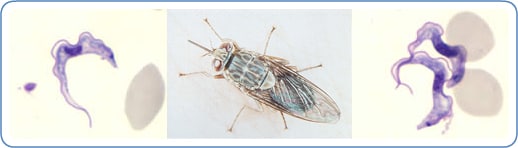
Transmission of T. brucei between mammal hosts is usually by an insect vector, the tsetse fly. T. brucei parasites undergo complex morphological changes as they move between insect and mammal over the course of their life cycle. The mammalian bloodstream forms are notable for their variable surface glycoprotein (VSG) coats, which undergo remarkable antigenic variation, enabling persistent evasion of host adaptive immunity and chronic infection. T. brucei is one of only a few pathogens that can cross the blood brain barrier. There is an urgent need for the development of new drug therapies as current treatments can prove fatal to the patient.
Whilst not historically regarded as T. brucei subspecies due to their different means of transmission, clinical presentation, and loss of kinetoplast DNA, genetic analyses reveal that T. equiperdum and T. evansi are evolved from parasites very similar to T. b. brucei, and are thought to be members of the brucei clade.
Infection: Trypanosomiasis

The insect vector for T. brucei is the tsetse fly (genus Glossina). The initial site of infection is the midgut of the fly (procyclic life cycle stage) and as the infection progresses it migrates via the proventriculus to the salivary glands where it attaches to the salivary gland surface (linked with a differentiation into the epimastigote life cycle stage). In the salivary glands some parasites detach and undergo adaptations (differentiation into the metacyclic life cycle stage) in preparation for injection to the mammalian host with the fly saliva on biting. In the mammal host the parasite lives within the bloodstream (slender bloodstream life cycle stage). Some parasites undergo adaptations (differentiation into the stumpy bloodstream life cycle stage) where it can reinfect the fly vector as it takes a blood meal after biting. In later stages of a T. brucei infection of a mammalian host the parasite may migrate from the bloodstream to also infect the lymph and cerebrospinal fluids.
In addition to the major form of transmission via the tsetse fly T. brucei may be transferred between mammals via bodily fluid exchange, such as by blood transfusion or sexual contact, although this is thought to be rare.
There are three different subspecies of T. brucei, which cause different variants of trypanosomiasis.
- T. brucei gambiense â€" Causes slow onset chronic trypanosomiasis in humans. Most common in central and western Africa, where humans are thought to be the primary reservoir.
- T. brucei rhodesiense â€" Causes fast onset acute trypanosomiasis in humans. Most common in southern and eastern Africa, where game animals and livestock are thought to be the primary reservoir.
- T. brucei brucei â€" Causes animal African trypanosomiasis, along with several other species of trypanosoma. T. b. brucei is not human infective due to its susceptibility to lysis by Trypanosome Lytic Factor-1 (TLF-1). However, as it is closely related to, and shares fundamental features with the human infective subspecies, T. b. brucei is used as a model for human infections in laboratory and animal studies.
Genetics of T. brucei subspecies

There are two subpopulations of T. b. gambiense that possesses two distinct groups that differ in genotype and phenotype. Group 2 is more akin to T. b. brucei than group 1 T. b. gambiense.
All T. b. gambiense are resistant to killing by a serum component â€" trypanosome lytic factor (TLF) of which there are two types: TLF-1 and TLF-2. Group 1 T. b. gambiense parasites avoid uptake of the TLF particles while those of group 2 are able to either neutralize or compensate for the effects of TLF.
In contrast T. b. rhodesiense is dependent upon the expression of a serum resistance associated (SRA) gene. This gene is not found in T. b. gambiense.
Cell structure
The structure of the cell is fairly typical of eukaryotes (see eukaryotic cell). All major organelles are seen, including the nucleus, mitochondria, endoplasmic reticulum, Golgi apparatus etc. Unusual features include the single large mitochondria with the mitochondrial DNA structure known as the kinetoplast, and its association with the basal body of the flagellum. The cytoskeleton is made up of microtubules. The cell surface of the bloodstream form features a dense coat of variable surface glycoproteins (VSGs) which is replaced by an equally dense coat of procyclins when the parasite differentiates into the procylic in the tsetse fly midgut.
Trypanosomatids show several different classes of cellular organisation of which two are adopted by Trypanosoma brucei at different stages of the life cycle:
- Epimastigote â€" Basal body anterior of nucleus, with a long flagellum attached along the cell body (in the salivary glands).
- Trypomastigote â€" Basal body posterior of nucleus, with a long flagellum attached along the cell body (in most other life cycle stages).
These names are derived from the Greek mastig- meaning whip, referring to the trypanosome's whip-like flagellum.
T. brucei is found as a trypomastigote in the slender, stumpy, procyclic and metacyclic forms. The procylic form differentiates to the proliferitive epimastigote form in the salivary glands of the insect. Unlike some other trypanosomatids, the promastigote and amastigote form do not form part of the T. brucei life cycle.
Genome
The genome of T. brucei is made up of:
- 11 pairs of large chromosomes of 1 to 6 megabase pairs.
- 3â€"5 intermediate chromosomes of 200 to 500 kilobase pairs.
- Around 100 minichromosomes of around 50 to 100 kilobase pairs. These may be present in multiple copies per haploid genome.
Most genes are held on the large chromosomes, with the minichromosomes carrying only VSG genes. The genome has been sequenced and is available online [1].
The mitochondrial genome is found condensed into the kinetoplast, an unusual feature unique to the kinetoplastea class. The kinetoplast and the basal body of the flagellum are strongly associated via a cytoskeletal structure.
VSG surface coat
Main section: VSG coat
The surface of the trypanosome is covered by a dense coat of Variable Surface Glycoprotein (VSG), which allows persistence of an infecting trypanosome population in the host. See below.
Cytoskeleton
The cytoskeleton is predominantly made up of microtubules, forming a subpellicular corset. The microtubules lie parallel to each other along the long axis of the cell, with the number of microtubules at any point roughly proportional to the circumference of the cell at that point. As the cell grows (including for mitosis) additional microtubules grow between the existing tubules, leading to semiconservative inheritance of the cytoskeleton. The microtubules are orientated + at the posterior and - at the anterior.
Microfilament and intermediate filaments do not play a role in the cytoskeleton.
Flagellar structure
The trypanosome flagellum has two main structures. It is made up of a typical flagellar axoneme which lies parallel to the paraflagellar rod, a lattice structure of proteins unique to the kinetoplastida, euglenoids and dinoflagellates.
The microtubules of the flagellar axoneme lie in the normal 9+2 arrangement, orientated with the + at the anterior end and the - in the basal body. The a cytoskeletal structure extends from the basal body to the kinetoplast. The flagellum is bound to the cytoskeleton of the main cell body by four specialised microtubules, which run parallel and in the same direction to the flagellar tubulin.
The flagellar function is twofold â€" locomotion via oscillations along the attached flagellum and cell body, and attachment to the fly gut during the procyclic phase.
VSG coat
The surface of the trypanosome is covered by a dense coat of ~5 x 10⁶ molecules of Variable Surface Glycoprotein (VSG). This coat enables an infecting T. brucei population to persistently evade the host's immune system, allowing chronic infection. The two properties of the VSG coat that allow immune evasion are:
- Shielding â€" the dense nature of the VSG coat prevents the immune system of the mammalian host from accessing the plasma membrane or any other invariant surface epitopes (such as ion channels, transporters, receptors etc.) of the parasite. The coat is uniform, made up of millions of copies of the same molecule; therefore the only parts of the trypanosome the immune system can 'see' are the N-terminal loops of the VSG that make up the coat.
- Periodic antigenic variation â€" the VSG coat undergoes frequent stochastic genetic modification â€" 'switching' â€" allowing variants expressing a new VSG coat to escape the specific immune response raised against the previous coat.
The cell surface of the bloodstream form features a dense coat of variable surface glycoproteins (VSGs) which is replaced by an equally dense coat of procyclins when the parasite differentiates into the procylic form in the tsetse fly midgut.
Antigenic variation
VSG is highly immunogenic, and an immune response raised against a specific VSG coat will rapidly kill trypanosomes expressing this variant. Antibody-mediated trypanosome killing can also be observed in vitro by a complement-mediated lysis assay. However, with each cell division there is a possibility that one or both of the progeny will switch expression to change the VSG variant that is being expressed. The frequency of VSG switching has been measured to be approximately 0.1% per division. As T. brucei populations can peak at a size of 1011 within a host this rapid rate of switching ensures that the parasite population is constantly diverse. A diverse range of coats expressed by the trypanosome population means that the immune system is always one step behind: it takes several days for an immune response against a given VSG to develop, giving the population time to diversify as individuals undergo further switching events. Reiteration of this process prevents extinction of the infecting trypanosome population, allowing chronic persistence of parasites in the host, enhancing opportunities for transmission. The clinical effect of this cycle is successive 'waves' of parasitaemia (trypanosomes in the blood).
VSG structure
VSG genes are hugely variable at the sequence level. However, different VSG variants have strongly conserved structural features, allowing them to perform a similar shielding function. VSGs are made up of a highly variable N terminal domain of around 300 to 350 amino acids, and a more conserved C terminal domain of around 100 amino acids. N-terminal domains dimerise to form a bundle of four alpha helices, around which hang smaller structural features. VSG is anchored to the cell membrane via a glycophosphatidylinositol (GPI) anchorâ€"a covalent linkage from the C-terminus, to approximately four sugars, to a phosphatidylinositol phospholipid acid which lies in the cell membrane.
VSG archive structure
The source of VSG variability during infection is a large 'archive' of VSG genes present in the T. brucei genome. Some of these are full-length, intact genes; others are pseudogenes) typically with frameshift mutations, premature stop codons, or fragmentation. Expression of an antigenically different VSG can occur by simply switching to a different full-length VSG gene. In addition, chimeric or 'mosaic' VSG genes can be generated by combining segments from more than one silent VSG gene. The formation of mosaic VSGs allows the (partial) expression of pseudogene VSGs, which can constitute the major portion of the VSG archive, and can contribute directly to antigenic variation, vastly increasing the trypanosome's capacity for immune evasion and potentially posing a major problem for vaccine development.
VSG expression
One major focus in trypanosome research is how all but one of the VSG genes are kept silent at a given time, and how these the active VSG is switched. The expressed VSG is always located in an Expression Site (ES), which are specialised expression loci found at the telomeres of some of the large and intermediate chromosomes. Each ES is a polycistronic unit, containing a number of Expression Site-Associated Genes (ESAGs) all expressed along with the active VSG. While multiple ES exist, only a single one is ever active at one time. A number of mechanisms appear to be involved in this process, but the exact nature of the silencing is still unclear.
The expressed VSG can be switched either by activating a different expression site (and thus changing to express the VSG in that site), or by changing the VSG gene in the active site to a different variant. The genome contains many copies of VSG genes, both on minichromosomes and in repeated sections in the interior of the chromosomes. These are generally silent, typically with omitted sections or premature stop codons, but are important in the evolution of new VSG genes. It is estimated up to 10% of the T.brucei genome may be made up of VSG genes or pseudogenes. Any of these genes can be moved into the active site by recombination for expression. Again, the exact mechanisms that control this are unclear, but the process seems to rely on DNA repair machinery and a process of homologous recombination.
Cell division

The mitotic division of T.brucei is unusual compared to most eukaryotes. The nuclear membrane remains intact and the chromosomes do not condense during mitosis. The basal body, unlike the centrosome of most eukaryotic cells, does not play a role in the organisation of the spindle and instead is involved in division of the kinetoplast.
Stages of mitosis:
- The basal body duplicates and both remain associated with the kinetoplast.
- Kinetoplast DNA undergoes synthesis then the kinetoplast divides coupled with separation of the two basal bodies.
- Nuclear DNA undergoes synthesis while a new flagellum extends from the younger, more posterior, basal body.
- The nucleus undergoes mitosis.
- Cytokinesis progresses from the anterior to posterior.
- Division completes with abscission.
Meiosis
Peacock et al. (2014) recently reported that T. brucei undergoes meiosis in its tsetse fly vector, and that meiosis is likely a normal part of the developmental cycle of T. brucei. They found that meiosis gives rise to haploid promastigote-like gametes that can interact with each other via their flagella and undergo cell fusion. Thus T. brucei is essentially a sexual organism. Trypanosomes belong to the supergroup Excavata and are one of the earliest diverging lineages among eukaryotes. Peacock et al. (2014) noted that their finding of a sexual stage in T. brucei supports the hypothesis that meiosis and sexual reproduction are ancestral and ubiquitous features of eukaryotes.
Killing by human serum and resistance to human serum killing
Trypanosoma brucei brucei (as well as related species T. equiperdum and T. evansi) is not human infective because it is susceptible to innate immune system 'trypanolytic' factors present in the serum of some primates, including humans. These trypanolytic factors have been identified as two serum complexes designated trypanolytic factors (TLF-1 and -2) both of which contain haptoglobin related protein (HPR) and apolipoprotein LI (ApoL1). TLF-1 is a member of the high density lipoprotein family of particles while TLF-2 is a related high molecular weight serum protein binding complex. The protein components of TLF-1 are haptoglobin related protein (HPR), apolipoprotein L-1 (apoL-1) and apolipoprotein A-1 (apoA-1). These three proteins are colocalized within spherical particles containing phospholipids and cholesterol. The protein components of TLF-2 include IgM and apolipoprotein A-I.
Trypanolytic factors are found only in a few species including humans, gorillas, mandrills, baboons and sooty mangabeys. This appears to be because haptoglobin related protein and apolipoprotein L-1 are unique to primates. This suggests these gene appeared in the primate genome 25 million years ago-35 million years ago.
Human infective subspecies T. b. gambiense and T. b. rhodesiense have evolved mechanisms of resisting the trypanolytic factors, described below.
ApoL1
ApoL1 is a member of a six gene family, ApoL1-6, that have arisen by tandem duplication. These proteins are normally involved in host apoptosis or autophagic death and possess a Bcl-2 homology domain 3. ApoL1 has been identified as the toxic component involved in trypanolysis. ApoLs have been subject to recent selective evolution possibly related to resistance to pathogens
The gene encoding ApoL1 is found on the long arm of chromosome 22 (22q12.3). Variants of this gene, termed G1 and G2, provide protection against T. b. rhodesiense. These benefits are not without their downside as a specific ApoL1 glomeropathy has been identified. This glomeropathy may help to explain the greater prevalence of hypertension in African populations.
The gene encodes a protein of 383 residues, including a typical signal peptide of 12 amino acids. The plasma protein is a single chain polypeptide with an apparent molecular mass of 42 kiloDaltons. ApoL1 has a membrane pore forming domain functionally similar to that of bacterial colicins. This domain is flanked by the membrane addressing domain and both these domains are required for parasite killing.
Within the kidney, ApoL1 is found in the podocytes in the glomeruli, the proximal tubular epithelium and the arteriolar endothelium. It has a high affinity for phosphatidic acid and cardiolipin and can be induced by interferon gamma and tumor necrosis factor alpha.
Hpr
Hpr is 91% identical to haptoglobin (Hp), an abundant acute phase serum protein, which possesses a high affinity for haemoglobin (Hb). When Hb is released from erythrocytes undergoing intravascular hemolysis Hp forms a complex with the Hb and these are removed from the circulation by the CD163 scavenger receptor. In contrast to Hpâ€"Hb, the Hprâ€"Hb complex does not bind CD163 and the Hpr serum concentration appears to be unaffected by haemolysis.
Killing mechanism
The association HPR with haemoglobin allows TLF-1 binding and uptake via the trypanosome haptoglobin-hemoglobin receptor (TbHpHbR). TLF-2 enters trypanosomes independently of TbHpHbR. TLF-1 uptake is enhanced in the low levels of haptoglobin which competes with haptoglobin related protein to bind free haemoglobin in the serum. However the complete absence of haptoglobin is associated with a decreased killing rate by serum.
The trypanosome haptoglobin-hemoglobin receptor is an elongated three a-helical bundle with a small membrane distal head. This protein extends above the variant surface glycoprotein layer that surrounds the parasite.
The first step in the killing mechanism is the binding of TLF to high affinity receptorsâ€"the haptoglobin-hemoglobin receptorsâ€"that are located in the flagellar pocket of the parasite. The bound TLF is endocytosed via coated vesicles and then trafficked to the parasite lysosomes. ApoL1 is the main lethal factor in the TLFs and kills trypanosomes after insertion into endosomal / lysosomal membranes. After ingestion by the parasite, the TLF-1 particle is trafficked to the lysosome wherein Apo1 is activated by a pH mediated conformational change. After fusion with the lysosome the pH drops from ~7 to ~5. This induces a conformational change in the ApoL1 membrane addressing domain which in turn causes a salt bridge linked hinge to open. This releases ApoL1 from the HDL particle to insert in the lysosomal membrane. The ApoL1 protein then creates anionic pores in the membrane which leads to depolarization of the membrane, a continuous influx of chloride and subsequent osmotic swelling of the lysosome. This influx in its turn leads to rupture of the lysosome and the subsequent death of the parasite.
Resistance mechanisms: T. b. gambiense
Trypanosoma brucei gambiense causes 97% of human cases of sleeping sickness. Resistance to ApoL1 is principally mediated by the hydrophobic ß-sheet of the T. b. gambiense specific glycoprotein. Other factors involved in resistance appear to be a change in the cysteine protease activity and TbHpHbR inactivation due to a leucine to serine substitution (L210S) at codon 210. This is due to a thymidine to cytosine mutation at the second codon position.
These mutations may have evolved due to the coexistence of malaria where this parasite is found. Haptoglobin levels are low in malaria because of the haemolysis that occurs with the release of the merozoites into the blood. The rupture of the erythrocytes results in the release of free haem into the blood where it is bound by haptoglobin. The haem is then removed along with the bound haptoglobin from the blood by the reticuloendothelial system.
Resistance mechanisms: T. b. rhodesiense
Trypanosoma brucei rhodesiense relies on a different mechanism of resistance: the serum resistance associated protein (SRA). The SRA gene is a truncated version of the major and variable surface antigen of the parasite, the variant surface glycoprotein. It has a low sequence homology with the VSGc (<25%). SRA is an expression site associated gene in T. b. rhodesiense and is located upstream of the VSGs in the active telomeric expression site. The protein is largely localized to small cytoplasmic vesicles between the flagellar pocket and the nucleus. In T. b. rhodesiense the TLF is directed to SRA containing endosomes while some dispute remain on its presence in the lysosome. SRA binds to ApoL1 using a coiledâ€"coiled interaction at the ApoL1 SRA interacting domain while within the trypanosome lysosome. This interaction prevents the release of the ApoL1 protein and the subsequent lysis of the lysosome and death of the parasite.
Baboons are known to be resistant to Trypanosoma brucei rhodesiense. The baboon version of the ApoL1 gene differs from the human gene in a number of respects including two critical lysines near the C terminus that are necessary and sufficient to prevent baboon ApoL1 binding to SRA. Experimental mutations allowing ApoL1 to be protected from neutralization by SRA have been shown capable of conferring trypanolytic activity on T. b. rhodesiense. These mutations resemble those found in baboons, but also resemble natural mutations conferring protection of humans against T. b. rhodesiense which are linked to kidney disease .


0 comments:
Post a Comment