
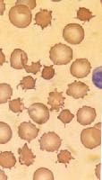
Acanthocyte (from the Greek word ἄκανθα acantha, meaning 'thorn'), in human biology and medicine, refers to a form of red blood cell that has a spiked cell membrane, due to abnormal thorny projections. A similar term is spur cells.
Acanthocytes have coarse, irregularly spaced, variably sized crenations, resembling many-pointed stars. They are seen on blood films in, among others abetalipoproteinemia, liver disease, chorea acanthocytosis, McLeod syndrome, and several inherited neurological disorders, such as neuroacanthocytosis, anorexia nervosa, infantile pyknocytosis, hypothyroidism, ideopathic neonatal hepatitis, alcoholism, congestive splenomegaly, Zieve syndrome, and chronic granulomatous disease.
Usage

Spur cells may refer synonymously to acanthocytes, or may refer in some sources to a specific subset of 'extreme acanthocytes' that have undergone splenic modification whereby additional cell membrane loss has blunted the spicules and the cells have become spherocytic ('spheroacanthocyte'), as seen in some patients with severe liver disease.
Acanthocytosis can refer generally to the presence of this type of crenated red blood cell, such as may be found in severe cirrhosis or pancreatitis, but can refer specifically to abetalipoproteinemia, a clinical condition with acanthocytic red blood cells, neurologic problems and steatorrhea. This particular cause of acanthocytosis (also known as abetalipoproteinemia, apolipoprotein B deficiency, and Bassen-Kornzweig syndrome) is a rare, genetically inherited, autosomal recessive condition due to the inability to fully digest dietary fats in the intestines as a result of various mutations of the microsomal triglyceride transfer protein (MTTP) gene.
Pathophysiology
Acanthocytes arise from either of two mechanisms. Alterations in membrane lipids are seen in abetalipoproteinemia and liver dysfunction. Alteration in membrane structural proteins are seen in neuroacanthocytosis and McLeod syndrome.
In liver dysfunction, apolipoprotein A-II deficient lipoprotein accumulates in plasma causing increased cholesterol in RBCs. This causes abnormalities of membrane of RBC causing remodeling in spleen and formation of acanthocytes.
In abetalipoproteinemia, there is deficiency of lipids and Vitamin E causing abnormal morphology of RBCs.
Differential diagnoses

Acanthocytosis can be seen in: Acute or Chronic Anemia, Hepatitis A,B,C; Hepatorenal syndrome, Hypopitutarism, Malabsorption Syndromes, Malnutrition In malnourishment, such as anorexia nervosa and cystic fibrosis, acanthocytosis remits with resolution of the nutritional deficiency. Acanthocyte-like cells may be found in hypothyroidism, after splenectomy, and in myelodysplasia.
Acanthocytes should be distinguished from echinocytes, which are also called 'burr cells', which although crenated are dissimilar in that they have multiple, small, projecting spiculations at regular intervals on the cell membrane. Burr cells usually imply uremia, but are seen in many conditions, including mild hemolysis in hypomagnesemia and hypophosphatemia, hemolytic anemia in long-distance runners, and pyruvate kinase deficiency. Burr cells can also arise in vitro due to elevated pH, blood storage, ATP depletion, calcium accumulation, and contact with glass. Acanthocytes should also be distinguished from keratocytes, also called 'horn cells' which have a few very large protuberances.


0 comments:
Post a Comment